本系列文章来自国外资料,本人对文章进行了初步翻译,采用中英文对照模式发布。还添加了更多的自己拍摄或检验同行拍摄的经典显微镜图片,以及临床诊断价值。
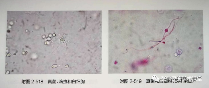
翻译及编辑这篇文章,是因为我曾经在许多年前见过这种细菌,因为不能准确识别,也曾经误认,并在我以前主编的三本尿液有形成分分析的专著也标出为真菌、不明真菌、酵母样真菌等名称,(下图),也描述过中间部位有球状膨起的形态。图 刊登在《实用尿液有形成分分析技术》第2版
后来有微生物专业的同行告诉我,这是一种用过抗生素的细菌的L形,我不置可否。今天看看外国同行是怎样研究和解释的,同时也希望纠正我之前的错误。本文以全英文原文和中文对照的方式呈现给大家。
• 原文:Post by Jose A. T. Poloni;Urine Sediment of the Month: Bacterial Variant FormsUrinary tract infections (UTI) are among the most common medical disorders in the world. Bacteria are the main infectious agent that causes UTIs, and bacteriuria and leukocyturia are the hallmarks of urinary sediment in patients with UTI. Bacteria can be observed in the urine sediment both as cocci and as rods, and Escherichia coli and Klebsiella pneumoniae as the most common bacteria causing UTI. Usually there is good correlation between the urine sediment findings and the results obtained by urine culture.
尿路感染(UTI)是世界上最常见的医学疾病之一。细菌是引起UTI的主要传染源,细菌尿和白细胞尿是UTI患者尿沉渣的特征。在尿沉渣中可以观察到球菌和杆菌,大肠杆菌和肺炎克雷伯菌是引起UTI的最常见细菌。通常尿沉渣检查结果与尿培养结果之间存在良好的相关性。However, urine microscopy can be limited by some false-positive scenarios (e.g.: urine contamination from genital secretions; bacterial overgrowth that can be observed when urine is not handled and analysed immediately after micturition) and false-negative scenarios (e.g.: cocci are less easily identifiable than rods, and some cocci can be confused with amorphous crystals.)
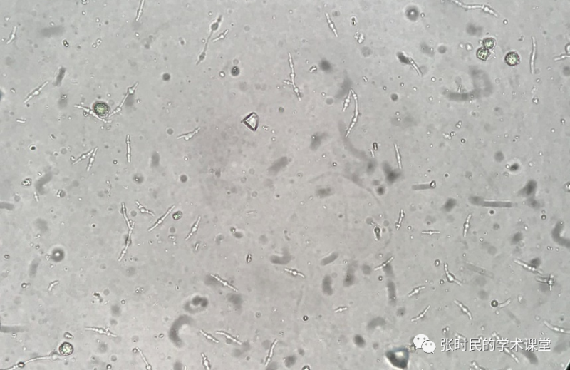
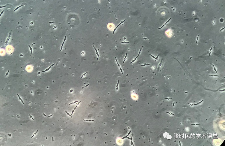
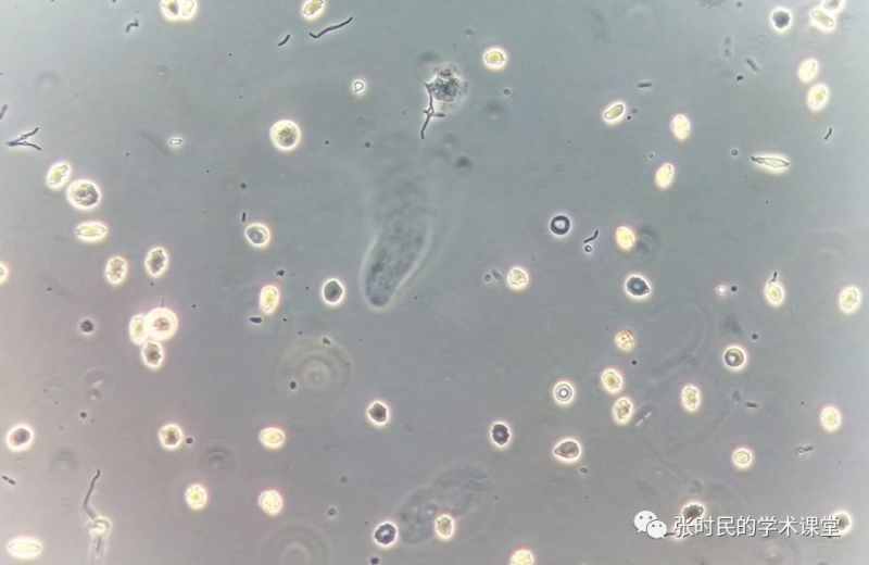
然而,尿液显微镜检查可能会出现一些假阳性情况(例如:生殖道分泌物导致的尿液污染;排尿后不立即处理导致分析尿液时可观察到细菌过度的生长)和假阴性情况(例如:球菌比杆状菌更难识别,一些球菌可能与无定形尿晶体混淆)的限制。Fig 1. Fresh and unstained urine sediment. Original magnification 400x. Bright field microscopy.图1 新鲜未染色的尿液沉渣。原图稿放大400倍。光学显微镜。Fig. 2 Fresh and unstained urine sediment. Original magnification 400x. Phase conrast microscopy.图2 新鲜和未染色的尿液沉渣。原图稿放大400倍。相位差显微镜。However, a bacterial variant form called “spheroplast” (Figures 1-6) has been described. It appears as an elongated structure with a round part in the middle of the “body” of the bacteria. It is a source of doubt for urine microscopists as itcan be misidentified as fungi or other structure. This challenge is not limited to the human eye: automated urinalysis systems can misidentify this form as an erythrocyte. It is not a common structure but due to the high chance of misidentification of the structures as other urinary elements, it is important to know how to properly identify of this kind of urinary particle.然而,一种名为“球形体”的细菌变异体在图1到图6中已经多次出现。它看起来是一个细长的结构,在菌体的“身体”中间有一个圆形的部分。这是尿液显微镜学家常常怀疑的一个问题,因为它可能被错误地识别为真菌或其他结构。这一挑战不仅限于人眼识别,自动尿液分析系统可能会错误地将这种形式的细菌识别为红细胞。这不是一种常见的结构,但由于这些结构与其他尿液成分很容易被错误识别,因此了解如何正确识别这种尿颗粒很重要。Fig. 3 Gram-stained urine sediment. Original magnification 1000x (oil immersion). Bright field microscopy.
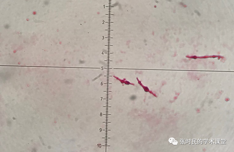
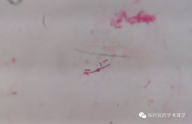
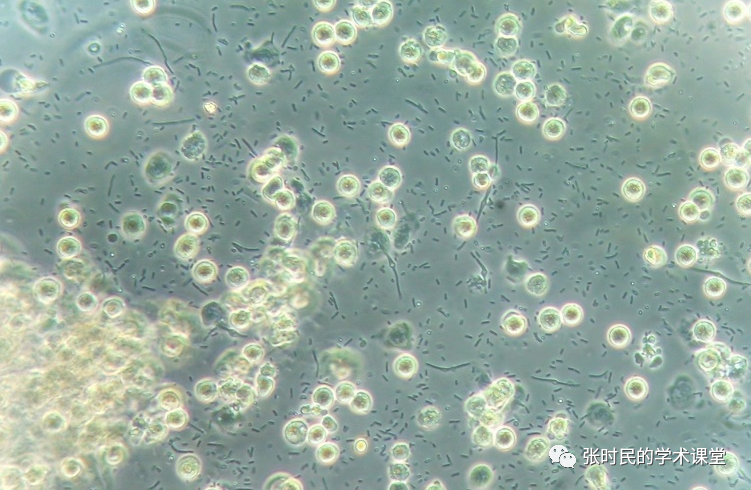
图3:革兰染色尿沉渣。原始放大倍率1000倍(油浸)。亮场显微镜。The bacterial shape is determined by the structure of its cell wall, which can be easily modified to survive environment conditions. The peptidoglycan and various cytoskeletal proteins located in the bacterial cell wall (e.g.: MreB, crescentin, FtsZ), play a major role in shaping the bacterial cells and changing their form. These abnormal bacterial forms are called cell wall deficient (CWD)-forms which have been classified as protoplasts and spheroplasts, depending on the degree of the loss of cell wall components. It has been observed that low concentrations of antibiotics can induce the change of bacterial form. For example, β-lactam antibiotics produce shape changes at concentrations significantly below those necessary to stop bacterial multiplication. The β-lactams act by binding to penicillin-binding proteins (PBPs) which induce the peptidoglycan synthesis. Subinhibitory concentrations of these antibiotics result in formation of abnormal, spherical forms that can be observed in the urine of treated patient.细菌的形状是由其细胞壁的结构决定的,细胞壁可以很容易地被改变,以适应环境的条件。位于细菌细胞壁上的肽聚糖和各种细胞骨架蛋白(例如:MreB、新月体、FtsZ)在塑造细菌细胞和改变其形态方面起着重要作用。这些异常的细菌形式被称为细胞壁缺陷型(CWD)——根据细胞壁成分的损失程度,可分为原生质体和球形体。据观察,低浓度的抗生素会引起细菌形态的改变。例如,β-内酰胺类抗生素在浓度显著低于阻止细菌增殖所需浓度时产生形状变化。β-内酰胺通过与青霉素结合蛋白(PBPs)结合而起作用,后者诱导肽聚糖的合成。这些抗生素的亚抑制浓度会导致在接受治疗的患者尿液中观察到异常的球形体。Fig. 4 Fresh and unstained urine sediment. Original magnification 400x. Phase contrast microscopy.图4:新鲜和未染色的尿液沉淀物。原图稿放大400倍。相衬显微镜。Fig. 5 Gram stained urine sediment. Original magnification 1000x (oil immersion). Bright field microscopy.图5:革兰染色尿沉渣。原始放大倍率1000倍(油浸)。光学显微镜。The real clinical significance of the observation of spheroplasts in the urine is not clear since the literature is extremely limited, however, in the few cases reported it is linked to resistant bacteria. Knowledge of this urinary particle is necessary avoid misidentification with other more common elements.在尿液中观察球形体的真正临床意义尚不清楚,因为文献报告极其有限,然而,在报告的少数病例中,它与耐药细菌有关。了解这种尿液颗粒是必要的,以避免与其他更常见的成分错误识别。Fig. 6 Fresh and unstained urine sediment. Original magnification 400x. Phase contrast microscopy.图6:新鲜和未染色的尿液沉淀物。原稿放大400倍。相衬显微镜。