• Post by Juan Carlos Velez
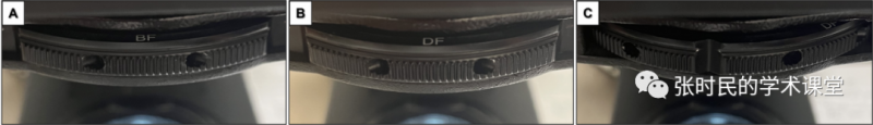
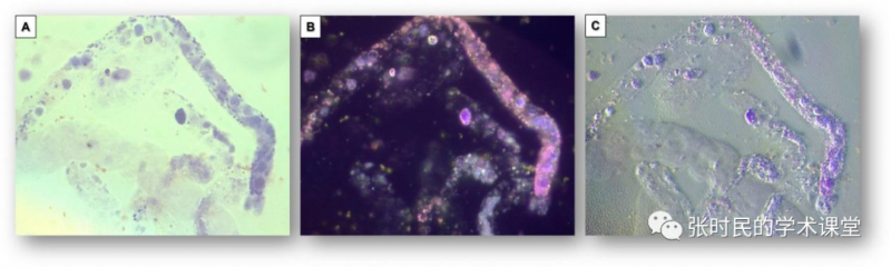
Urinary sediment microscopy is a diagnostic technique designed to acquire 2-dimensional images of structures present in a slide. However, 2-dimensional views may lack sufficient contrast to allow for optimal visualization. One way to enhance the images and create a 3-dimensional view is by applying oblique illumination.尿沉渣显微镜检查是一种诊断技术,旨在获取载玻片中结构的二维图像。然而,二维视图可能缺乏足够的对比度,无法实现最佳可视化。增强图像并创建三维视图的一种方法是应用倾斜照明。By allowing the light to project onto the field through only 1 side of the condenser, structures cast a shadow and create a 3-D-like image. Standard microscopes do not routinely come from the manufacturer set up to provide this type of illumination unless they get customized. However, oblique illumination can be easily achieved by taking advantage of the artifactual effect of partially rotating the condenser diaphragm turret between 2 fixed settings.使光仅通过聚光镜的一侧投射到视场上,尿液有形成分的结构投射出阴影,并创建类似三维的图像。除非进行定制,否则标准显微镜通常不会提供此类照明。然而,通过利用在两个固定设置之间部分,人工旋转聚光镜振膜转台的效果,可以轻松实现倾斜照明。Figure 1. Condenser turret illustrating settings at (A) bright field (BF) and (B) dark field (DF). By rotating the turret ¾ of the way between BF and DF (C), oblique illumination can be achieved.图1:聚光镜转台,说明(A)亮视野(BF);(B)暗视野(DF)的设置;通过BF和DF(C)之间旋转盘¾位置,可以实现倾斜照明。The 2 approaches that offer the best balance of light and contrast to generate a “relief” or “satellite view” effect are by rotating the condenser turret between bright field and dark field illumination (Figure 1) or between phase contrast and bright field illumination setting.提供最佳光线和对比度平衡以产生“浮雕”或“卫星视图”效果的两种方法是通过在亮场和暗场照明(图1)之间或在相位对比度和亮场照明设置之间旋转聚光镜转台。Figure 2. Bilirubin-stained renal tubular epithelial cell casts (RTECC) under bright field microscopy (A and C). The cast matrix is better visualized under oblique illumination (B and D) Images at 400x magnification.图2:亮视野显微镜下胆红素染色的肾小管上皮细胞管型(A和C)。在400倍放大的倾斜照明(B和D)图像下,投射矩阵更好地可视化。Figure 3. White blood cell cast under bright (A) and dark field (B) illumination (Sternheimer-Malbin stain). While the cast matrix is noticeable along half of the cast (right side of the images), it is difficult to visualize its full length. Oblique illumination (C) allows better distinction of the cast matrix. In addition, other hyaline casts (some containing cells) become more apparent. Images obtained at 400x magnification.Oblique illumination can be particularly helpful to visualize a cast matrix that may not be very visible on bright or dark field illumination, particularly when phase contrast microscopy is not available (Figures2-4).图3:在明视野(A)和暗视野场(B)照明下,白细胞管型(Sternheimer-Malbin染色)。虽然投射矩阵沿投射的一半(图像的右侧)很明显,但很难看到其全长。倾斜照明(C)后可以更好地区分投射矩阵。此外,其他透明管型(一些含有细胞)变得更加明显,放大400倍获得的图像。
倾斜照明特别有助于可视化在明场或暗场照明下可能不太可见的铸造基质,尤其是在没有相衬显微镜的情况下(图2-4)。
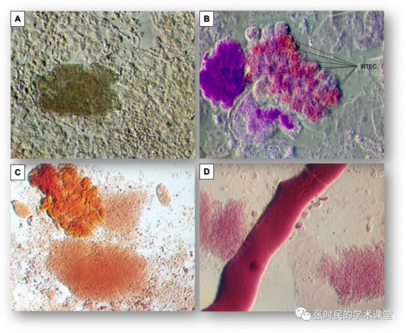
Figure 4. Oblique illumination accentuates (A) the granularity of coarse granular casts. Yeast (pseudo-hyphae) can be more noticeable; (B) nuclei from renal tubular epithelial cells (RTEC) can be nicely outlined; texture of broad (C) cracked and (D) smooth waxy casts becomes more visible.
It also allows for crisp visualization of cell nuclei and thus better characterization of the type of cells or cells free-floating in the urine sediment or embedded within casts.
图4:倾斜照明突出了(A)粗颗粒管型的粒度。酵母(假菌丝)更明显;(B) 肾小管上皮细胞(RTEC)的细胞核轮廓清晰;宽(C)裂纹和(D)光滑蜡样管型的纹理变得更加明显。
它还可以清晰地显示细胞核,从而更好地表征细胞类型,或游离在尿液沉积物中,或嵌入管型内的细胞。
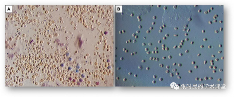
Figure 5. Dysmorphic erythrocyturia, scattered acanthocytes and leukocytes from a patient with crescentic glomerulonephritis (A). Crenated erythrocytes from a urine specimen contaminated due to metrorrhagia (B). Squamous epithelial cells are also seen in the upper side of the image. As seen in both panels, red blood cell morphology can be clearly appreciated under oblique illumination.
The characteristic dysmorphism of acanthocytes is also nicely revealed by oblique illumination (Figure 5).
图5:新月体肾炎患者尿中的畸形红细胞,散在的棘细胞和白细胞(A)。由于子宫出血而被污染的尿液样本中的皱缩红细胞(B)。鳞状上皮细胞也可见于图像的上部。如两幅图所示,在倾斜照明下可以清楚地观察到红细胞的形态。· 译者注:倾斜照明就是在多功能显微镜的聚光器旋转台上,选择一个两种模式的一个中间位置,以达到特殊模拟3D效果的图像构成,是一种尝试。
编辑:骆秉涵